One test could change your life.
The right testing tells you what’s wrong. We tell you how to fix it.
One test could change everything
Most people just haven't had the right one.
Not All Lab Work Is Equal
The testing method your clinic uses determines how accurate your results are. Most clinics use the cheapest option available, and when your numbers are off, every decision that follows is based on bad information.
For hormones like testosterone and estradiol, accuracy depends on the testing method. Most clinics use immunoassay because it is cheaper and faster. It is also less reliable at the exact ranges where treatment decisions are being made. LC-MS/MS is the methodology used in clinical research, and it is what we use.

Inflated Panel Counts
Some clinics advertise 100+ biomarkers but pad the number with calculated values derived from other results. A calculated marker is not a test. It is a math equation applied to a test you already paid for, and most clinics do not disclose which markers are measured or estimated.

Tech Company or Clinic?
The biggest names in this space are tech companies with a health feature. Their product is a dashboard, an app, a health score. The clinical relationship, if it exists at all, is an add-on. A dashboard does not evaluate your symptoms, interpret your labs in context, or adjust your treatment when something changes. A medical professional does.

Why This Matters To You.
These are not just statistics and are only a fraction of health issues men face. They represent a current and accelerating crisis affecting every aspect of male health.
The data from 2022-2025 shows these trends are worsening, not improving.
"You have a choice. Accept the statistics and become another data point, or reject 'normal for your age' and take control of your health."Start Protocol
More than markers
A Live Consultation With A Licensed Medical Provider
Your first panel includes a video consultation with a licensed medical provider who reviews your results with you and explains what they mean in the context of your health and your goals.
Access to Treatment
Your blood work opens access to our full prescribing formulary. Testosterone, GLP-1 medications, peptides, and more. If your labs indicate that treatment is appropriate, your provider can prescribe during the same consultation.
No Subscriptions or Membership Plans
You pay for your panel and your treatment. There are no monthly memberships, no recurring charges, and no platform fees. When you need follow-up labs or a refill, you order what you need.
Every marker, categorized.
Browse by body system. Filter by tier to see exactly what you get on each panel.
Hematology Blood 9 markers Markers describing red and white cell counts and oxygen-carrying capacity.
Hematocrit
Also known as Bioavaiable
The unbound fraction
True hormone status Treatment Response Symptom correlation
Red Blood Cells
The total number of erythrocytes circulating in peripheral blood, each containing hemoglobin for oxygen transport.
The concentration of red blood cells per unit volume of blood.
Elevated levels (erythrocytosis/polycythemia) indicate chronic hypoxia, dehydration, polycythemia vera, high altitude adaptation, smoking, or erythropoietin-secreting tumors. Decreased levels indicate anemia from blood loss, nutritional deficiency (iron, B12, folate), hemolysis, bone marrow suppression, or chronic disease RBC count can be influenced by altitude, hydration status, pregnancy, testosterone levels, and certain medications (erythropoietin, anabolic steroids).
Hemoglobin
The iron-containing protein within red blood cells responsible for binding and transporting oxygen from the lungs to tissues and returning carbon dioxide to the lungs.
The concentration of hemoglobin in whole blood, reflecting oxygen-carrying capacity.
Elevated levels indicate chronic hypoxia, dehydration, polycythemia vera, high altitude, smoking, or erythropoietin-secreting tumors. Decreased levels (anemia) indicate blood loss, nutritional deficiency (iron, B12, folate), hemolysis, bone marrow suppression, chronic kidney disease, or chronic inflammation. Hemoglobin can be influenced by altitude, hydration status, pregnancy, age, ethnicity (lower in African Americans), and smoking.
Mean corpuscular volume (MCV)
A calculated red blood cell index that measures the average size and volume of red blood cells. MCV is calculated from the hematocrit percentage divided by the red blood cell count.
The average volume of individual red blood cells, used to classify anemias into three categories: microcytic (MCV 100 fL).
Large red blood cells suggest vitamin B12 deficiency, folate deficiency, excessive alcohol use, liver disease, or certain medications. Small red blood cells suggest iron deficiency, thalassemia (an inherited blood disorder), chronic disease, or lead exposure. MCV increases slightly with age; reticulocytes (young red blood cells) are larger and can raise the number; combined deficiencies (iron plus B12) can produce a falsely normal MCV because the effects cancel each other out..
Mean corpuscular hemoglobin (MCH)
A calculated red blood cell index that measures the average amount of hemoglobin in each red blood cell.
The average mass of hemoglobin per red blood cell, calculated by dividing total hemoglobin by the red blood cell count. MCH correlates closely with MCV.
Elevated levels often correspond with macrocytic anemia (larger than normal red blood cells) caused by Vitamin B12 or folate deficiency, liver disease, alcohol abuse, or an underactive thyroid. Low levels (hypochromia) typically indicate iron deficiency anemia or thalassemia, where red blood cells are smaller than normal and lack sufficient hemoglobin. MCH status can be falsely elevated by hyperlipidemia (high blood fats), very high white blood cell counts, or the presence of cold agglutinins (antibodies that cause red blood cells to clump together).
Mean Corpuscular Hemoglobin Concentration (MCHC)
A calculated value that represents the average density or concentration of hemoglobin in a given volume of packed red blood cells
The density of hemoglobin within each red blood cell. Unlike MCH (which measures total hemoglobin mass per cell), MCHC accounts for cell size by relating hemoglobin content to cell volume.
Elevated levels (hyperchromia) are rare and physiologically limited but can indicate Hereditary Spherocytosis. Levels may also appear falsely high due to cold agglutinins (clumped cells) or lipemia (fatty blood). Low MCHC indicates hypochromic red blood cells (paler than normal when viewed under microscope). Can also indicate Iron deficiency anemia, thalassemia, anemia of chronic disease, sideroblastic anemia, lead poisoning. MCHC status can be falsely elevated by autoagglutination (cells clumping in the tube), high cholesterol/triglycerides, or hemolysis (broken cells).
Red cell distribution width (RDW)
A calculated value from your complete blood count that measures the degree of variation in the size of your red blood cells.
The coefficient of variation of the red blood cell volume distribution, essentially measuring how consistent or inconsistent the red blood cells are in size.
Elevated levels indicate significant heterogeneity in cell size, commonly seen in early nutritional deficiencies (iron, B12, or folate) before anemia becomes overt, hemolytic anemia, liver disease, or following recent blood transfusions. Low levels indicate that red blood cells are very uniform in size; while often normal, this can also occur in chronic anemias where cells are consistently small (microcytic) or large (macrocytic) rather than mixed. RDW status can be influenced by a high reticulocyte count (immature red blood cells are larger), alcohol abuse, and the fragmentation of cells due to heart valves or severe infection.
Platelet Count
Platelets (also called thrombocytes) are small, colorless blood cell fragments produced in the bone marrow and are essential for normal blood clotting and wound healing
The number of platelets per microliter of blood, assessing the body's ability to form clots and stop bleeding
Elevated levels (thrombocytosis) can indicate chronic inflammation, infection, iron deficiency anemia, removal of the spleen (splenectomy), or myeloproliferative disorders like essential thrombocythemia Low levels (thrombocytopenia) indicate a risk of excess bleeding and may result from bone marrow suppression (chemotherapy, radiation), autoimmune destruction (ITP), viral infections (Hepatitis C, HIV), or chronic liver disease. Platelet status can be influenced by recent surgery, physical exertion, acute infection (which often causes a temporary rise), and alcohol consumption (which can lower counts).
Mean Platelet Volume (MPV)
A measurement of the average size of platelets in the blood, which can reflect platelet age and bone marrow activity
Platelet size. Larger platelets are usually younger and more active. Smaller platelets tend to be older.
Elevated levels suggest the bone marrow is rapidly producing new platelets, often seen in immune thrombocytopenia (ITP) where platelets are destroyed and replaced quickly, or in recovery from chemotherapy. Low levels indicate older average platelets, typically seen when the bone marrow is underproducing (aplastic anemia) or suppressed by cytotoxic drugs. MPV status can be influenced by the time between blood draw and analysis (platelets swell in EDTA tubes over time) and cardiovascular risk factors like obesity and diabetes.
Cardio-metabolic Insight Cardiovascular 20 markers Markers that help frame lipid balance, inflammation, and cardiovascular risk.
Cholesterol, Total
Cholesterol is a waxy, fat-like substance essential for cell membrane structure, hormone synthesis, vitamin D production, and bile acid formation.
The sum of all cholesterol carried in lipoproteins: LDL (low-density lipoprotein), HDL (high-density lipoprotein), and VLDL (very low-density lipoprotein).
Elevated levels may indicate familial or polygenic hypercholesterolemia, diet high in saturated/trans fats, hypothyroidism, nephrotic syndrome, primary biliary cirrhosis, obstructive liver disease, or diabetes mellitus. Low levels may indicate hyperthyroidism, severe liver disease, malnutrition, malabsorption, chronic anemia, sepsis, critical illness, inflammatory conditions, or inherited disorders (abetalipoproteinemia, hypobetalipoproteinemia, Tangier disease). Cholesterol may be influenced by both Testosterone and Estrogen levels.
HDL Cholesterol
High-Density Lipoprotein Cholesterol is often called "good" cholesterol because HDL particles transport cholesterol from peripheral tissues and arterial walls back to the liver for excretion or recycling
The concentration of cholesterol carried within high-density lipoprotein particles.
Elevated levels are desirable and are associated with a reduced risk of heart disease and stroke ("cardioprotective"). Low levels may indicate sedentary lifestyle, obesity, smoking, type 2 diabetes, metabolic syndrome, insulin resistance, hypertriglyceridemia, chronic kidney disease, malnutrition, certain medications (beta-blockers, anabolic steroids, progestins), or genetic conditions. Cholesterol may be influenced by both Testosterone and Estrogen levels.
Triglycerides
Triglycerides are the most abundant lipids in the body, serving as the primary form of stored energy. They are synthesized from dietary fats and excess carbohydrates.
The amount of fat circulating in the bloodstream.
Elevated levels may indicate excess caloric intake (especially refined carbohydrates and alcohol), obesity, poorly controlled diabetes mellitus, metabolic syndrome, hypothyroidism, nephrotic syndrome, chronic kidney disease, primary biliary cirrhosis, or familial hypertriglyceridemia. Low levels may indicate malnutrition, malabsorption, hyperthyroidism, abetalipoproteinemia, or very low-fat diets. Low triglycerides are generally not clinically concerning. Inadequate fasting is the most common cause of spuriously elevated results.
LDL Cholesterol
Low-Density Lipoprotein Cholesterol is the primary carrier of cholesterol to peripheral tissues.
The cholesterol content within LDL particles. Most commonly calculated using the Friedewald equation (Total Cholesterol − HDL-C − Triglycerides/5).
Elevated levels may indicate familial hypercholesterolemia, polygenic hypercholesterolemia, diet high in saturated/trans fats, hypothyroidism, nephrotic syndrome, obstructive liver disease, diabetes mellitus, obesity, or medications (thiazides, cyclosporine, glucocorticoids). Low levels may indicate hyperthyroidism, malnutrition, malabsorption, severe liver disease, sepsis, critical illness, or inherited disorders (abetalipoproteinemia, hypobetalipoproteinemia). Nonfasting samples may underestimate LDL
Total Cholesterol to HDL Ratio
A calculated score that compares your total cholesterol to your "good" (HDL) cholesterol.
The proportion of total cholesterol relative to HDL cholesterol.
Elevated ratio may indicate low HDL cholesterol, elevated LDL or VLDL cholesterol, or a combination. Conditions associated with elevated ratio include metabolic syndrome, obesity, diabetes, sedentary lifestyle, smoking, and familial dyslipidemias. An unfavorable ratio signals increased cardiovascular risk even when individual lipid values appear borderline. Low ratio may indicate high HDL (exercise, genetic factors, moderate alcohol intake) relative to total cholesterol, or very low LDL from aggressive lipid-lowering therapy. Calculated from total cholesterol and HDL values; accuracy depends on accuracy of component measurements.
LDL/HDL Ratio
This ratio compares atherogenic LDL cholesterol to HDL cholesterol.
The proportion of LDL cholesterol relative to HDL cholesterol.
Elevated ratio may indicate elevated LDL, low HDL, or both. Associated with increased atherogenic burden and cardiovascular risk. Conditions elevating this ratio mirror those affecting its components: familial hypercholesterolemia, metabolic syndrome, diabetes, obesity, hypothyroidism, sedentary lifestyle, smoking. Low ratio may indicate well-controlled LDL (through lifestyle or medication), naturally high HDL, or both. Calculated from LDL and HDL values. When triglycerides exceed calculation thresholds for LDL, the ratio becomes unreliable unless direct LDL measurement is used.
Non HDL Cholesterol
The combined cholesterol content of all non-HDL lipoproteins (Total Cholesterol − HDL-C).
Represents the total cholesterol content of all atherogenic lipoproteins: LDL, VLDL, IDL, and lipoprotein(a).
Elevated levels may indicate elevated LDL, elevated VLDL (often from hypertriglyceridemia), elevated IDL, elevated lipoprotein(a), or combinations thereof. Associated conditions parallel those affecting LDL: familial hyperlipidemia, metabolic syndrome, diabetes, obesity, hypothyroidism, nephrotic syndrome, and atherogenic diets. Low levels may indicate hyperthyroidism, malnutrition, malabsorption, severe liver disease, or aggressive lipid-lowering therapy. Calculated from total cholesterol and HDL; accuracy depends on component measurements.
HDL P
LDL particle number is a measurement of the total concentration of low-density lipoprotein particles circulating in the bloodstream, determined via nuclear magnetic resonance (NMR) spectroscopy, and represents the actual count of atherogenic particles rather than their cholesterol content.
Serum LDL-P concentration reflects the total number of apoB-containing LDL particles per liter, with each particle containing exactly one apolipoprotein B molecule, providing a direct measure of atherogenic particle burden that may identify cardiovascular risk even when LDL cholesterol appears normal.
Elevated levels may indicate increased atherosclerotic cardiovascular disease risk, insulin resistance, metabolic syndrome, familial hypercholesterolemia, or discordance between LDL-C and true atherogenic burden. Low levels are generally favorable and associated with reduced cardiovascular risk. LDL-P can be influenced by diet, triglyceride levels, insulin resistance, medications (statins, fibrates, niacin), thyroid function, and genetic factors affecting lipid metabolism.
HDL SIZE
HDL size measures the average diameter of high-density lipoprotein particles in nanometers, reflecting the degree of lipid enrichment and maturation of the HDL particle population.
Average HDL particle diameter reflects the predominant stage of HDL particle maturation, with larger particles typically indicating greater cholesterol ester content and more advanced reverse cholesterol transport capacity.
Larger HDL particles may reflect more mature, cholesterol-enriched particles, though clinical significance varies. HDL particles may indicate nascent, less mature particles or impaired HDL maturation. HDL size can be influenced by exercise, alcohol consumption, triglyceride levels, genetic factors (CETP activity), and medications.
LARGE HDL P
Large HDL particle number quantifies the concentration of larger, more mature HDL particles that have accumulated substantial cholesterol ester content through the reverse cholesterol transport process.
Serum concentration of large HDL particles reflects the population of fully matured HDL capable of delivering cholesterol to the liver for excretion.
Higher concentrations of large HDL particles may reflect efficient reverse cholesterol transport and favorable lipid metabolism. Reduced large HDL-P may indicate impaired HDL maturation or accelerated HDL catabolism. Large HDL-P can be influenced by exercise, alcohol consumption, and lipid-modifying medications.
LARGE VLDL P
Large VLDL particle number quantifies the concentration of triglyceride-enriched VLDL particles, which are associated with atherogenic remnant lipoproteins and insulin resistance.
Serum concentration of large VLDL particles reflects hepatic triglyceride secretion and the population of triglyceride-rich lipoproteins that may contribute to cardiovascular risk through remnant formation.
Elevated large VLDL-P may indicate insulin resistance, metabolic syndrome, or elevated cardiovascular risk from atherogenic remnant particles. Lower concentrations are generally favorable. Large VLDL-P can be influenced by dietary intake (particularly refined carbohydrates and alcohol), fasting status, insulin sensitivity, and lipid-modifying medications.
LDL P
LDL particle number is a measurement of the total concentration of low-density lipoprotein particles circulating in the bloodstream and represents the actual count of atherogenic particles rather than their cholesterol content.
Serum LDL-P concentration reflects the total number of apoB-containing LDL particles per liter, with each particle containing exactly one apolipoprotein B molecule, providing a direct measure of atherogenic particle burden that may identify cardiovascular risk even when LDL cholesterol appears normal.
Elevated levels may indicate increased atherosclerotic cardiovascular disease risk, insulin resistance, metabolic syndrome, familial hypercholesterolemia, or discordance between LDL-C and true atherogenic burden. Low levels are generally favorable and associated with reduced cardiovascular risk. LDL-P can be influenced by diet, triglyceride levels, insulin resistance, medications (statins, fibrates, niacin), thyroid function, and genetic factors affecting lipid metabolism.
LDL SIZE
LDL size is a measurement of the average diameter of LDL particles expressed in nanometers, determined via NMR spectroscopy, distinguishing between larger, more buoyant particles and smaller, denser particles.
Average LDL particle diameter reflects the predominant phenotype of circulating LDL, with smaller, dense LDL (pattern B) traditionally associated with elevated cardiovascular risk, though this relationship becomes less predictive after accounting for total LDL particle number.
Larger particle size (>21.2 nm) is generally associated with pattern A phenotype and considered more favorable, though particle number remains the stronger predictor of cardiovascular events. Smaller particle size (<20.5 nm) indicates pattern B phenotype, often associated with insulin resistance, elevated triglycerides, and metabolic syndrome. LDL size can be influenced by triglyceride levels, insulin resistance, dietary carbohydrate intake, genetic factors, and medications affecting lipid metabolism.
SMALL LDL P
Small LDL particle number quantifies the concentration of smaller, denser LDL particles within the total LDL particle population, measured via NMR spectroscopy.
Serum concentration of small LDL particles reflects the atherogenic subset of LDL particles that may penetrate arterial walls more readily, though this metric's independent predictive value diminishes when total LDL-P is taken into account.
Elevated levels may indicate insulin resistance, metabolic syndrome, elevated triglycerides, or pattern B LDL phenotype associated with cardiovascular risk. Lower concentrations of small LDL particles are generally favorable. Small LDL-P can be influenced by dietary carbohydrate intake, triglyceride levels, insulin sensitivity, weight status, and lipid-modifying medications.
VLDL SIZE
VLDL size measures the average diameter of very-low-density lipoprotein particles, which are triglyceride-rich lipoproteins synthesized by the liver and serve as precursors to LDL particles.
Average VLDL particle diameter reflects the triglyceride content and metabolic processing status of these particles, with larger VLDL particles typically containing more triglycerides.
Larger VLDL particles may indicate elevated hepatic triglyceride secretion, often associated with insulin resistance or metabolic syndrome. Smaller VLDL particles may reflect lower triglyceride content or efficient lipolysis. VLDL size can be influenced by dietary fat and carbohydrate intake, fasting status, insulin sensitivity, alcohol consumption, and lipid-modifying medications.
Homocysteine
Homocysteine is a sulfur-containing amino acid intermediate produced during methionine metabolism, whose clearance requires adequate B6, B12, and folate for remethylation and transsulfuration pathways.
Serum homocysteine concentration reflects the efficiency of methionine metabolism and serves as a functional marker of B-vitamin status (B6, B12, folate), with elevations indicating pathway impairment.
Elevated levels may indicate B6, B12, or folate deficiency, MTHFR gene polymorphisms, chronic kidney disease, hypothyroidism, or elevated cardiovascular and cerebrovascular disease risk. Low homocysteine is generally not clinically concerning. Homocysteine status can be influenced by B-vitamin intake, renal function, age (increases with age), coffee consumption, certain medications, MTHFR polymorphisms, creatine intake, and dietary methionine intake.
Apolipoprotein A1
Apolipoprotein A1 (Apo A-1) is the principal structural protein of high-density lipoprotein (HDL), constituting approximately 70% of HDL protein content.
Serum Apo A-1 concentration reflects the protein component responsible for HDL's atheroprotective functions, including cholesterol efflux from peripheral tissues and delivery to the liver for excretion.
Higher Apo A-1 levels are generally associated with favorable cardiovascular profiles; very high levels may occur with familial hyperalphalipoproteinemia, estrogen therapy, or statin use. Low levels may indicate increased cardiovascular disease risk, Tangier disease (rare genetic HDL deficiency), familial hypoalphalipoproteinemia, liver disease, nephrotic syndrome, uncontrolled diabetes, or smoking. Apo A-1 status can be influenced by sex (higher in females), exercise (increases levels), alcohol consumption, smoking (decreases levels), estrogen status, and lipid-modifying medications.
Apolipoprotein B
Apolipoprotein B (Apo B) is the essential structural protein of atherogenic lipoproteins, with exactly one Apo B molecule per LDL, VLDL, IDL, and Lp(a) particle, making it a direct measure of total atherogenic particle number.
Serum Apo B concentration represents the total number of potentially atherogenic particles in circulation, providing a single measurement that captures all particles capable of penetrating arterial walls and initiating atherosclerosis.
Elevated levels indicate increased atherogenic particle burden and cardiovascular risk, familial hypercholesterolemia, metabolic syndrome, or type 2 diabetes; superior to LDL-C for risk assessment when discordance exists. Low levels are generally favorable; very low levels may occur with hypobetalipoproteinemia or abetalipoproteinemia (rare genetic disorders). Apo B status can be influenced by diet, weight status, insulin resistance, statin therapy (decreases levels), and genetic factors affecting lipoprotein metabolism.
Apolipoprotein B/A1 Ratio
The Apo B/Apo A-1 ratio integrates the balance between atherogenic particles (Apo B) and anti-atherogenic HDL particles (Apo A-1) into a single metric that may provide superior cardiovascular risk stratification compared to traditional lipid ratios.
This ratio reflects the balance between pro-atherogenic and anti-atherogenic lipoproteins, with higher ratios indicating greater atherogenic potential and lower ratios indicating more favorable lipid profiles.
Elevated Apo B/Apo A-1 ratio indicates unfavorable balance between atherogenic and protective lipoproteins, associated with increased cardiovascular disease risk and abdominal aortic aneurysm risk. Lower ratios are associated with reduced cardiovascular risk. This ratio can be influenced by lifestyle factors, medications, and genetic factors affecting both Apo B and Apo A-1 metabolism; may detect risk 20 years before cardiovascular events.
hs CRP
High-sensitivity C-reactive protein (hs-CRP) is an acute-phase reactant synthesized by the liver in response to inflammatory cytokines, with the high-sensitivity assay capable of detecting subtle elevations associated with low-grade systemic inflammation and cardiovascular risk.
The general level of inflammation in the body, specifically used to assess cardiovascular risk.
Elevated levels in the cardiovascular risk range (>3.0 mg/L) may indicate increased cardiovascular event risk, residual inflammatory risk despite statin therapy, metabolic syndrome, or chronic low-grade inflammation; values >10 mg/L suggest acute infection, injury, or active inflammatory disease. Low levels (<1.0 mg/L) are associated with lower cardiovascular risk. hs-CRP can be influenced by acute infections, chronic inflammatory conditions (rheumatoid arthritis, IBD), obesity, smoking, diabetes, estrogen therapy, or statin use (decreases levels.
Electrolyte Balance Electrolytes 5 markers Markers governing fluid balance, nerve, and muscle function.
Sodium
Sodium is an electrolyte that plays a central role in maintaining fluid balance, blood pressure, and proper nerve and muscle function
Sodium concentration in blood, reflecting the balance between sodium intake, fluid intake, and kidney excretion.
Elevated levels (hypernatremia) almost always indicate dehydration (loss of free water) or high salt intake; rare causes include diabetes insipidus or kidney dysfunction. Low levels (hyponatremia) can be caused by overhydration, diuretic use, heart failure, liver disease, or hormonal imbalances. Sodium and water balance are tightly linked. Changes in sodium concentration usually reflect fluid balance rather than true sodium deficiency or excess.
Potassium
Potassium is an electrolyte essential for muscle contraction (including heart muscle), nerve transmission, and moving nutrients into cells.
The concentration of potassium in the blood.
Elevated levels (hyperkalemia) are dangerous and can cause cardiac arrhythmias; causes include kidney failure, cell damage (burns/trauma), or certain blood pressure medications (ACE inhibitorsARBs, potassium-sparing diuretics, beta-blockers). Low levels (hypokalemia) can cause muscle weakness and heart palpitations; common causes are diuretic use, chronic diarrhea, or vomiting. Potassium levels can be falsely elevated if the blood sample is "hemolyzed" (red blood cells burst during the draw), releasing their potassium into the serum.
Chloride
Chloride is an electrolyte that works with sodium and potassium to maintain fluid balance, blood pressure, and acid-base balance.
Chloride concentration in blood.
Elevated levels (hyperchloremia) often mimic sodium elevations (dehydration) or can indicate metabolic acidosis (loss of bicarbonate). Low levels (hypochloremia) are frequently caused by prolonged vomiting (loss of stomach acid/HCl) or metabolic alkalosis. Chloride levels generally mirror sodium levels; if sodium goes up, chloride usually follows.
Carbon Dioxide
Carbon dioxide in blood is measured as bicarbonate, which your body produces as a waste product of metabolism. Bicarbonate acts as a buffer to maintain the acid-base balance (pH) of your blood
The acid-base status of your blood
Elevated levels typically indicate metabolic alkalosis (too much base), often due to severe vomiting or lung diseases that cause CO2 retention (like COPD). Low levels indicate metabolic acidosis (too much acid), which can be seen in kidney disease, diabetic ketoacidosis, or toxicity (aspirin/methanol).
Calcium
The most abundant mineral in the body, essential for bone health, muscle contraction, and nerve transmission.
The total amount of calcium in the blood, including both the free (ionized) calcium and calcium bound to proteins like albumin.
Elevated levels (hypercalcemia) are most commonly caused by hyperparathyroidism (overactive parathyroid glands) or malignancy (cancer). Low levels (hypocalcemia) can result from Vitamin D deficiency, kidney failure, hypoparathyroidism, or low albumin levels. Since much of the calcium is carried by albumin, low protein levels can make calcium appear low even if the active (ionized) calcium is normal.
Hormonal Balance Hormones 10 markers Markers tracking endocrine signalling across reproductive and stress hormones.
Total Testosterone (Uncapped, LCMS)
Testosterone is a hormone which regulates sex drive, erectile function, sperm production, muscle mass, bone density, red blood cell production, fat distribution, and mood.
The total concentration of testosterone in blood, reflecting testicular function in men and ovarian/adrenal androgen production in women.
Low levels may indicate primary hypogonadism (testicular failure from Klinefelter syndrome, testicular injury, infection, chemotherapy, or radiation), secondary hypogonadism (pituitary or hypothalamic dysfunction from tumors, Kallmann syndrome, or hyperprolactinemia), or late-onset/adult-onset hypogonadism associated with aging, obesity, type 2 diabetes, or chronic illness. Elevated levels may indicate androgen-secreting tumors of the adrenal glands or testes, congenital adrenal hyperplasia, hyperthyroidism (which increases SHBG and may elevate total testosterone), or exogenous androgen use (testosterone replacement therapy, anabolic steroids). In women, elevated testosterone suggests polycystic ovary syndrome (PCOS), ovarian tumors, adrenal tumors, or congenital adrenal hyperplasia. Immunoassays (what most clinics use) are prone to cross-reactivity with structurally similar steroids and can produce results that are 20% lower or up to five-fold higher than actual values, particularly at low concentrations found in women, children, and hypogonadal men. LC-MS/MS separates testosterone from similar compounds before quantifying it by molecular mass, eliminating interference and providing reliable results across the entire measurable range.
Free Testosterone (Dialysis, not calculated)
Free testosterone is the unbound fraction of testosterone circulating in blood (typically less than 2–3% of total testosterone). Only the free fraction can enter cells and exert biological effects, making it the "active" form.
The actual amount of "bioavailable" hormone currently ready to work on the body's tissues (muscle, brain, libido).
Elevated levels often indicate exogenous testosterone use (TRT), where the sheer volume of hormone saturates the carrier proteins, leaving more floating free. In women, it is a key marker for Polycystic Ovary Syndrome (PCOS). Low levels are the most accurate indicator of true hypogonadism (Low T symptoms). A man can have "normal" Total Testosterone but feel terrible because high SHBG (a carrier protein) is locking it all up, leaving his Free Testosterone critically low. Specimen collection should occur in early morning. Different methods produce different reference ranges that are not directly comparable.
DHEA - S
The sulfated ester of DHEA, produced almost exclusively by the adrenal cortex and serving as the most abundant circulating steroid hormone which functions primarily as a reservoir for conversion to androgens and estrogens in peripheral tissues.
The serum concentration of DHEA-S, reflecting adrenal androgen production and functional adrenal reserve.
Elevated levels in women can cause acne, hair loss, and facial hair growth (PCOS); in men, it typically causes no symptoms but may indicate adrenal tumors. Low levels are associated with aging ("adropause"), adrenal insufficiency, low libido, fatigue, and depression. DHEA-S status can be influenced by age (declines annually after age 30), sex, chronic stress, insulin resistance, and medications including corticosteroids and insulin sensitizers.
SHBG
Sex Hormone Binding Globulin is a hepatically synthesized glycoprotein that binds and transports testosterone and estradiol in circulation, thereby regulating the bioavailability of these sex steroids to target tissues.
he capacity of the blood to bind sex hormones; this determines how much of your total testosterone is actually free to work.
Elevated levels result in less free testosterone; causes include hyperthyroidism, liver disease, low-protein diets, estrogen therapy and aging. Low levels result in more free testosterone but faster clearance; causes include obesity, Type 2 diabetes, hypothyroidism, and high-sugar diets, or androgen use. SHBG status can be influenced by thyroid function, insulin sensitivity, liver health, estrogen/androgen balance, medications, and genetic polymorphisms.
PSA Total
Prostate-Specific Antigen is a protein produced by prostate cells used to screen for prostate conditions.
The total concentration of prostate-specific antigen in serum, including both free and protein-bound fractions.
Elevated levels may indicate Benign Prostatic Hyperplasia (BPH), infection (prostatitis), or prostate cancer. TRT can cause mild elevations. Low levels are normal; however, 5-alpha reductase inhibitors (Finasteride/Dutasteride) artificially lower results by ~50%. PSA status can be influenced by age, prostate volume, medications (5-alpha reductase inhibitors), recent sexual activity, prostate manipulation, and infection or inflammation.
Estradiol (Ultra Sensitive, LCMS)
The most potent and abundant estrogen in humans, produced primarily by the ovarian follicles in premenopausal women and through peripheral aromatization of testosterone in men and postmenopausal women. Estradiol regulates the menstrual cycle, maintains bone density, supports cardiovascular and cognitive function, and influences libido and mood in both sexes.
The serum concentration of estradiol (E2) with high precision across the full physiological range, including the low levels found in men, children, and postmenopausal women.
High levels in men cause gynecomastia, fluid retention, and emotional volatility; in women, it indicates the ovulation phase or estrogen dominance. Low levels in men cause joint pain, osteoporosis, and low libido; in women, it indicates menopause or amenorrhea, and also osteoporosis.. Estradiol status can be influenced by body fat percentage, liver function, aromatase activity, medications (aromatase inhibitors, SERMs), menstrual cycle phase in premenopausal women, and time of day.
LH
Luteinizing Hormone is a gonadotropin secreted by the anterior pituitary that stimulates testosterone production by testicular Leydig cells in men and triggers ovulation while supporting corpus luteum function in women. It is essential for evaluating the hypothalamic-pituitary-gonadal axis and distinguishing primary from secondary hypogonadism
The serum concentration of luteinizing hormone, reflecting pituitary gonadotropin output in response to GnRH stimulation and gonadal feedback.
Elevated levels indicate Primary hypogonadism (testicular failure, premature ovarian insufficiency), menopause, polycystic ovary syndrome (PCOS—elevated LH:FSH ratio), Klinefelter syndrome, or anorchia. Low levels indicate Secondary (central) hypogonadism, pituitary or hypothalamic dysfunction, hyperprolactinemia, anabolic steroid use, severe illness or stress, excessive exercise. LH status can be influenced by pulsatile secretion (single measurements may not reflect true baseline), menstrual cycle phase (midcycle surge), medications (GnRH agonists/antagonists, opioids), and time of day.
FSH
Follicle-Stimulating Hormone is a gonadotropin secreted by the anterior pituitary that drives follicular development and estrogen production in women while stimulating Sertoli cells and spermatogenesis in men. Combined with LH, FSH is essential for distinguishing primary gonadal failure from hypothalamic-pituitary causes and assessing ovarian reserve in women.
The serum concentration of follicle-stimulating hormone, reflecting pituitary gonadotropin output and gonadal feedback status.
High levels indicate primary hypogonadism (ovarian failure, testicular failure), menopause, diminished ovarian reserve, Klinefelter syndrome, Turner syndrome, premature ovarian insufficiency. Low levels indicate secondary (central) hypogonadism, pituitary or hypothalamic dysfunction, hyperprolactinemia, anabolic steroid use, severe illness, excessive exercise, PCOS (relatively low compared to LH). FSH status can be influenced by menstrual cycle phase, age, medications (oral contraceptives, GnRH modulators), and pulsatile secretion.
Progesterone (Ultrasensitive, LCMS)
Progesterone is a steroid hormone produced primarily by the corpus luteum following ovulation and by the placenta during pregnancy, responsible for preparing the uterine lining for embryo implantation and maintaining early pregnancy. In men, progesterone is produced in small amounts by the adrenal glands and testes, serving as a precursor in steroid hormone synthesis pathways.
The serum concentration of progesterone with high specificity, reflecting corpus luteum function, ovulatory status, or pregnancy maintenance. The ultrasensitive method avoids cross-reactivity with DHEA and other steroids that can cause falsely elevated immunoassay results,
High levels indicate ovulation confirmed (luteal phase), pregnancy, ovarian cysts, congenital adrenal hyperplasia, certain ovarian tumors. Low levels can indicate anovulation, luteal phase deficiency (associated with recurrent miscarriage), threatened miscarriage, ectopic pregnancy. Progesterone status can be influenced by menstrual cycle timing, medications (progestins, fertility treatments), and adrenal function.
IGF-1
Insulin-like growth factor 1 is a polypeptide hormone structurally similar to insulin that mediates many growth hormone (GH) effects, playing essential roles in tissue growth, metabolic regulation, and cellular proliferation throughout life.
Serum IGF-1 concentration reflects integrated growth hormone secretion over time, as IGF-1 has a longer half-life and more stable levels than pulsatile GH, making it the preferred marker for assessing GH axis status.
Elevated levels may indicate acromegaly (GH-secreting pituitary tumor), excessive GH replacement therapy, or gigantism in children; very high levels require evaluation for underlying GH excess. Low levels may indicate growth hormone deficiency (childhood or adult-onset), pituitary dysfunction, malnutrition, anorexia, liver disease, or uncontrolled diabetes; levels below the 2.5th percentile for age (Z-score < -2) suggest GH deficiency. IGF-1 status can be influenced by age (peaks in adolescence, declines thereafter), nutritional status, liver function, obesity (may paradoxically lower levels), thyroid status, and medications.
Immune & Inflammation Immune 11 markers Markers reflecting immune cell counts and underlying inflammatory tone.
White Blood Cells
The total number of leukocytes circulating in peripheral blood, comprising neutrophils, lymphocytes, monocytes, eosinophils, and basophils.
The concentration of all white blood cells reflecting overall immune system activity.
Elevated levels (leukocytosis) indicate infection, inflammation, physical or emotional stress, tissue damage, allergic reactions, corticosteroid use, or hematologic malignancy. Decreased levels (leukopenia) indicate bone marrow suppression, viral infection, autoimmune disease, overwhelming sepsis, or medication effects (chemotherapy, certain antibiotics). WBC counts can be influenced by time of day, exercise, pregnancy, smoking, ethnicity (lower baseline in individuals of African and Middle Eastern descent), and splenectomy.
Absolute Neutrophils
A calculated measurement reflecting the total number of neutrophils circulating in the blood
How many neutrophils are available to fight infection. Calculated by multiplying total white blood cell count by the percentage of neutrophils.
Elevated levels (neutrophilia) typically indicate an active bacterial infection, acute stress (physical or emotional), tissue necrosis (such as from a heart attack or burn), or chronic inflammation (like rheumatoid arthritis). Medications such as corticosteroids can also cause significant elevation. Low levels (neutropenia) indicate a weakened immune system and increased susceptibility to infection. This can be caused by viral infections (like flu or hepatitis), autoimmune disorders, chemotherapy, severe vitamin deficiencies (B12/folate), or bone marrow failure. Neutrophil status can be influenced by recent intense exercise, smoking, pregnancy, and "demargination" (where stress causes cells sticking to blood vessel walls to release into the stream, temporarily raising the count).
Absolute Lymphocytes
A calculated value showing the total number of lymphocytes in your blood.
The actual number of lymphocyte cells per microliter of blood, derived by multiplying the total white blood cell count by the percentage of lymphocytes
Elevated levels (lymphocytosis) typically indicate an acute viral infection (such as Epstein-Barr/Mono, Cytomegalovirus, or Hepatitis), chronic bacterial infections (like Pertussis), or chronic lymphocytic leukemia (CLL). Low levels (lymphopenia) indicate a compromised immune system, often caused by corticosteroid use, severe stress (trauma/surgery), malnutrition (zinc deficiency), autoimmune lupus, HIV infection, or following radiation therapy. Lymphocyte status can be heavily influenced by stress (which lowers counts rapidly via cortisol), fasting or malnutrition, and recent intense physical exercise (which can transiently elevate counts).
Absolute Monocytes
A calculated value showing the total number of monocytes in your blood.
The actual number of monocyte cells per microliter of blood, derived by multiplying the total white blood cell count by the percentage of monocytes.
Elevated levels (monocytosis) often indicate a chronic infection (such as tuberculosis or subacute bacterial endocarditis), recovery from an acute infection, autoimmune disease (like lupus or rheumatoid arthritis), or certain blood disorders (like chronic myelomonocytic leukemia). Low levels (monocytopenia) are rare in isolation but can occur during acute stress, severe infection (sepsis), or bone marrow suppression from chemotherapy or hairy cell leukemia. Monocyte status can be influenced by chronic inflammatory conditions and is often elevated during the recovery phase of an acute infection ("the cleanup crew" coming in after the neutrophils).
Absolute Eosinophils
A calculated value showing the total number of eosinophils in your blood.
The actual number of Eosinophils cells per microliter of blood.Calculated by multiplying total white blood cell count by the percentage of eosinophils.
Elevated levels (eosinophilia) most commonly indicate an allergic reaction (asthma, hay fever, drug allergy) or a parasitic infection (worms). It can also be seen in certain autoimmune diseases (vasculitis), adrenal insufficiency (Addison’s disease), or Hodgkin lymphoma. Low levels (eosinopenia) are associated with Cushing's syndrome (excess cortisol), acute bacterial infections, sepsis, corticosteroid use, stress response. Eosinophil status exhibits a diurnal variation (counts are lower in the morning due to higher cortisol levels) and can be significantly elevated by eczema or asthma flares.
Absolute Basophils
A calculated value showing the total number of basophils in your blood.
The actual number of basophil cells per microliter of blood. Calculated by multiplying total white blood cell count by the percentage of basophils.
Elevated levels (basophilia) are uncommon but can indicate chronic inflammation (ulcerative colitis), hypothyroidism, or myeloproliferative neoplasms (like chronic myeloid leukemia or polycythemia vera). Low levels (basopenia) are generally not clinically significant because the normal range includes zero, but can be associated with acute allergic reactions (where they degranulate and disappear from view) or hyperthyroidism. Basophils show diurnal variation where they are lowest in the morning and highest at night. Basophilia rarely occurs in isolation; it usually accompanies eosinophilia or other white blood cell abnormalities
Neutrophils
Neutrophils are white blood cells that serve as the body's primary defense against bacterial and fungal infections
The relative percentage of neutrophils compared to the total number of white blood cells in circulation.
Elevated levels (relative neutrophilia) typically indicate an active bacterial infection, acute inflammation (such as appendicitis or rheumatic fever), tissue necrosis (heart attack, burns), or significant physical or emotional stress. Low levels (relative neutropenia) are often seen in viral infections (flu, hepatitis), where lymphocytes increase and "crowd out" the neutrophil percentage, or in bone marrow suppression from chemotherapy or radiation. Neutrophil percentage can be temporarily elevated by "demargination," where stress or exercise causes cells sticking to vessel walls to release into the bloodstream, and by corticosteroid use.
Lymphocytes
Lymphocytes are white blood cells central to the immune system. There are three main types: T cells (attack infected and cancerous cells), B cells (make antibodies), and natural killer cells (destroy virus-infected and cancer cells).
The percentage of the total white blood cell count that consists of lymphocytes.
Elevated levels (relative lymphocytosis) are most commonly caused by viral infections (such as Mono, Flu, or Hepatitis), but can also indicate chronic bacterial infections or lymphocytic leukemia. Low levels (relative lymphopenia) typically occur when neutrophils are elevated (due to bacterial infection or stress), or can result from steroid use, autoimmune disorders like lupus, or HIV infection. Lymphocyte percentage is inversely related to neutrophil percentage
Monocytes
The largest type of white blood cell, which works to remove cellular debris and fights certain chronic infections.
The percentage of the total white blood cell count that consists of monocytes.
Elevated levels (relative monocytosis) often indicate the recovery phase of an acute infection, chronic inflammatory diseases, tuberculosis, or viral infections like measles or mumps. Low levels are generally not clinically significant in isolation but can occur during acute stress or severe infection. Can also appear low if other white cell types are elevated. Monocyte percentage often rises as neutrophil levels fall during recovery from an illness.
Eosinophils
Eosinophils are white blood cells that contain granules filled with toxic proteins used to kill parasites and respond to allergic reactions.
The percentage of the total white blood cell count that consists of eosinophils.
Elevated levels (relative eosinophilia) strongly suggest an allergic response (asthma, hay fever, drug allergy) or a parasitic infection. Low levels are difficult to detect due to the low baseline but may be caused by acute stress or corticosteroid use. osinophil percentage typically follows a daily rhythm, being lower in the morning when cortisol levels are higher.
Basophils
Basophils are the rarest white blood cells. They contain granules filled with histamine and heparin, which they release during allergic reactions and to help prevent blood clots.
The percentage of the total white blood cell count that consists of basophils.
Elevated levels (relative basophilia) are rare but can be seen in chronic inflammation, hypothyroidism, or certain bone marrow disorders. A reading of 0% is common on routine blood tests and rarely concerning because basophils are so few in number, but can be caused due to acute infections, severe allergic reactions (anaphylaxis), hyperthyroidism, corticosteroid use, or acute stress Basophils show diurnal variation, meaning lowest in the morning, highest at night. Basophilia rarely occurs alone; it usually accompanies eosinophilia or other white cell abnormalities.
Renal Function Snapshot Kidney 4 markers Markers that estimate filtration, waste handling, and hydration balance.
Urea Nitrogen (BUN)
Blood urea nitrogen is a waste product created when your liver breaks down protein. Urea nitrogen travels through your bloodstream to your kidneys, which filter it out and excrete it in urine.
How well your kidneys are filtering waste from your blood. Rising BUN suggests kidneys may not be removing waste efficiently.
Elevated levels (azotemia) typically indicate impaired kidney function, dehydration, a high-protein diet, gastrointestinal bleeding, or heart failure (due to reduced blood flow to the kidneys). Low levels are less common but can be seen in severe liver disease (where the liver cannot make urea), malnutrition (low protein intake), or overhydration. BUN status can be influenced by diet (high protein increases it) and hydration (dehydration concentrates it, raising the number).
Creatinine
Creatinine is a waste product generated from the normal breakdown of creatine in muscle tissue.
Kidney filtration function. Creatinine is freely filtered by the kidneys and not reabsorbed, making it a more stable marker of kidney function than BUN.
Elevated levels indicate that the kidneys are not filtering waste efficiently, suggesting kidney damage, failure, or blockage (kidney stones, prostate enlargement). Low levels are generally not a health concern but are typically associated with low muscle mass (common in elderly patients) or severe malnutrition. Creatinine status can be influenced by muscle mass (bodybuilders naturally have higher baselines) and certain supplements (like creatine monohydrate), which can artificially elevate levels without kidney damage.
Glomerular filtration rate (GFR)
A calculated estimate of how well your kidneys filter blood. The glomeruli are tiny clusters of blood vessels in the kidneys that remove waste products
The actual flow rate of filtered fluid through the kidney, serving as an overall index of kidney function.
Significantly high levels can occur in early pregnancy or early stages of diabetes (hyperfiltration), and in those with high protein intakes. Low levels indicate reduced kidney function; chronic kidney disease. eGFR is an estimate and may be less accurate in people with very high or very low muscle mass
BUN/Creatinine Ratio
A calculated ratio comparing blood urea nitrogen to creatinine. Because the kidneys handle these two waste products differently, the ratio helps distinguish between causes of abnormal kidney markers.
Whether kidney abnormalities are due to reduced blood flow to the kidneys (prerenal), intrinsic kidney damage (renal), or obstruction after the kidneys (postrenal).
Elevated ratios (>20:1) typically suggest dehydration, heart failure, or GI bleeding (where BUN rises disproportionately to creatinine). Low ratios may indicate liver disease or malnutrition (low urea production) or rhabdomyolysis (muscle breakdown causing high creatinine). This ratio helps differentiate between dehydration (high ratio) and actual kidney disease (normal ratio with both markers elevated).
Hepatic Function Panel Liver 8 markers Markers reflecting liver enzyme activity and metabolic clearance.
Protein, Total
A combined measurement of the two main types of protein in the blood: albumin and globulin.
Overall protein status, reflecting liver synthetic function, nutritional status, and immune system activity.
Elevated levels can indicate chronic inflammation or infection (high globulins) like Hepatitis C or HIV, or bone marrow disorders like Multiple Myeloma. Low levels often signal liver disease (reduced production), kidney disease (loss through urine), or malnutrition/malabsorption. Dehydration can falsely elevate total protein by concentrating the blood.
Albumin
Albumin is the most abundant protein in blood plasma, made by the liver. It keeps fluid from leaking out of blood vessels into tissues and carries hormones, vitamins, enzymes, and medications throughout your body.
The concentration of albumin, serving as a primary marker of liver synthetic function and nutritional status.
Elevated levels are almost exclusively caused by dehydration (hemoconcentration). Low levels indicate liver disease (cirrhosis), kidney syndrome (nephrotic syndrome), chronic inflammation, or severe malnutrition. Albumin has a long half-life (~20 days), so it reflects chronic rather than acute nutritional status.
Globulin
Globulins are a group of proteins made by the liver and immune system. They include alpha and beta globulins (transport proteins) and gamma globulins (immunoglobulins/antibodies).
Immune system activity and certain aspects of liver function. The calculated difference between Total Protein and Albumin.
Elevated levels often indicate chronic infection, autoimmune disease (Lupus, RA), or malignancies like Multiple Myeloma. Low levels can be a sign of renal disease or immune deficiency. Globulin is not usually measured directly but calculated; specific electrophoresis is needed to determine exactly which globulin fraction is high.
Albumin/Globulin Ratio
A calculated ratio comparing albumin levels to globulin levels in blood.
The balance between albumin and globulin production, reflecting liver function, immune activity, and overall protein status.
High ratios may indicate underproduction of immunoglobulins (hypogammaglobulinemia), certain leukemias, genetic immunodeficiencies, severe dehydration with high albumin. Low ratios suggest either low albumin (liver disease) or high globulin (chronic inflammation/infection), often serving as a nonspecific marker for systemic illness.
Bilirubin, Total
Bilirubin is a yellow pigment produced when red blood cells break down. The liver processes bilirubin, attaches it to a sugar molecule (conjugation), and excretes it in bile.
Liver function, bile duct patency, and red blood cell breakdown. Total bilirubin includes unconjugated (indirect) and conjugated (direct) forms.
Elevated levels (jaundice) indicate liver damage (hepatitis, cirrhosis), bile duct obstruction (gallstones), or excessive breakdown of red blood cells (hemolysis). Low levels are generally not a clinical concern. Gilbert syndrome affects 3–7% of the population and causes isolated, mild unconjugated hyperbilirubinemia that increases with fasting or illness but requires no treatment.
Alkaline Phosphatase
Alkaline phosphatase is an enzyme found primarily in liver, bone, kidneys, and intestines.
The activity of the ALP enzyme in the blood, reflecting liver and biliary tract function, as well as bone activity.
Elevated levels typically indicate bile duct obstruction (cholestasis) or active bone formation/turnover (such as in growing children, bone fractures, or Paget's disease). Low levels are rare but can indicate malnutrition (zinc/magnesium deficiency) or a genetic condition called hypophosphatasia. Elevated ALP requires determining whether the source is liver or bone.
Aspartate Aminotransferase (AST)
AST is an enzyme found in liver cells, heart muscle, skeletal muscle, and other tissues. When these tissues are damaged, AST is released into the bloodstream.
The level of AST enzyme, serving as a marker for cellular injury.
Elevated levels indicate liver damage (hepatitis, alcohol abuse, cirrhosis) or muscle damage (strenuous exercise, injury). Low levels are not clinically significant but can be associated with Vitamin B6 deficiency. Intense exercise can cause significant spikes in AST without liver disease.
Alanine transaminase (ALT)
ALT is an enzyme found primarily in liver cells. Because ALT is more concentrated in the liver than other tissues, it is a more specific marker of liver injury than AST.
Liver cell (hepatocyte) injury. When liver cells are damaged, ALT leaks into the bloodstream.
Elevated levels are a direct indicator of liver cell damage, commonly seen in fatty liver disease, hepatitis, alcohol abuse, or drug toxicity. Low levels are generally normal. ALT can be mildly elevated from vigorous exercise.
Metabolic Health Metabolic 5 markers Markers tied to insulin sensitivity, glycemic control, and energy metabolism.
Glucose
Glucose is a sugar that serves as your body's primary energy source. It comes mainly from carbohydrates in food.
The amount of sugar circulating in your bloodstream. Fasting glucose reflects baseline blood sugar when you haven't eaten for at least 8 hours.
Elevated levels (hyperglycemia) most commonly indicate diabetes (Type 1 or Type 2) or pre-diabetes, but can also be caused by acute stress, pancreatitis, hyperthyroidism, or corticosteroid use. Low levels (hypoglycemia) can result from excessive insulin dosage (in diabetics), starvation, liver disease, adrenal insufficiency, or rare insulin-secreting tumors (insulinomas). Blood glucose fluctuates throughout the day based on food intake, physical activity, and stress. Medications including corticosteroids, thiazides, beta-blockers, and certain psychiatric drugs can raise glucose levels.
Insulin
Insulin is a hormone produced by pancreatic beta cells that regulates blood glucose by facilitating cellular uptake of glucose into muscle, fat, and liver.
The amount of insulin circulating in the blood while fasting.
Elevated levels (hyperinsulinemia) are the earliest warning sign of insulin resistance and pre-diabetes, often rising years before blood sugar (glucose) or A1C become abnormal. The pancreas is "shouting" to keep blood sugar stable. Low levels indicate that the pancreas is struggling to produce enough insulin (Type 1 Diabetes or late-stage Type 2 Diabetes). Being in a non fasted state alters insulin levels.
Hemoglobin A1c
A form of hemoglobin (the oxygen-carrying protein in red blood cells) that has sugar attached to it. Because red blood cells live for about 3 months, measuring this provides a long-term record of average blood sugar levels.
Your average blood sugar level over the past 2–3 months.
Elevated levels indicate chronic high blood sugar, significantly increasing the risk of cardiovascular disease, nerve damage, kidney failure, and vision loss. Low levels are rare but can indicate recent severe blood loss, chronic anemia, or liver disease. Results can be misleading in people with anemia or recent blood transfusions.
Uric Acid
Uric acid is the terminal breakdown product of purine metabolism in humans, produced primarily by the liver and intestinal mucosa when the body degrades purines from dietary sources and cellular turnover.
The concentration of uric acid in the blood.
Elevated levels may indicate gout or gouty arthritis, uric acid kidney stones, metabolic syndrome, chronic kidney disease, conditions with increased cell turnover (leukemia, lymphoma, chemotherapy), excessive dietary purine intake, diuretic use, dehydration, or cardiovascular disease risk. Low levels may indicate Wilson's disease, Fanconi syndrome, certain medications (high-dose aspirin, allopurinol, probenecid), severe liver disease, high-dose vitamin C supplementation, malnutrition with low purine/protein intake, or rare genetic disorders affecting purine metabolism. Uric acid status can be influenced by age, sex, body weight, alcohol consumption, fructose intake, kidney function, certain medications (thiazide diuretics, cyclosporine), recent dietary purine consumption, and genetic factors.
Cortisol, Total
Cortisol is the primary glucocorticoid hormone produced by the adrenal cortex, essential for stress response, glucose metabolism, immune modulation, and circadian rhythm regulation, with secretion controlled by the hypothalamic-pituitary-adrenal (HPA) axis.
Serum total cortisol concentration reflects both protein-bound (~90–95%) and free cortisol, with levels exhibiting pronounced diurnal variation where it is highest in early morning and declining throughout the day.
Elevated levels may indicate Cushing syndrome (adrenal adenoma, carcinoma, pituitary ACTH-secreting tumor, or ectopic ACTH production), acute physiological stress, obesity, depression, hyperthyroidism, or exogenous corticosteroid use. Low levels may indicate primary adrenal insufficiency (Addison disease), secondary adrenal insufficiency (pituitary dysfunction), tertiary insufficiency (hypothalamic dysfunction), or abrupt discontinuation of chronic corticosteroid therapy. Cortisol levels can be influenced by collection time (diurnal variation), acute stress, sleep disruption, estrogen status (oral contraceptives, pregnancy increase binding protein), chronic illness, and medications.
Nutrient Status Nutrients 19 markers Markers that reveal how vitamin and mineral stores are tracking.
Vitamin D2
The liver metabolite of vitamin D2, a form of vitamin D that comes exclusively from plant/fungal dietary sources or supplements.
The concentration of vitamin D2 circulating in the blood, which reflects recent dietary intake of D2-fortified foods or ergocalciferol supplementation.
Elevated levels indicate excessive D2 supplementation and may contribute to toxicity if total vitamin D is significantly raised. In unsupplemented individuals, undetectable D2 is normal and expected; in patients on D2 therapy, low levels may indicate malabsorption. Vitamin D2 status can be influenced by malabsorption disorders, liver disease, and medications such as anticonvulsants.
Vitamin D3
The liver metabolite of cholecalciferol (vitamin D3), the primary form of vitamin D in humans, produced endogenously in the skin upon ultraviolet B exposure or obtained from animal-derived foods and supplements.
The serum concentration of 25-hydroxyvitamin D3, reflecting the combination of sun exposure, dietary intake from animal sources (fatty fish, egg yolks, liver), and D3 supplementation.
Elevated levels indicate excessive supplementation or sun exposure and may contribute to hypercalcemia, hypercalciuria, and soft tissue calcification if total vitamin D exceeds safe thresholds. Low levels suggest inadequate sun exposure, poor dietary intake, malabsorption, liver dysfunction, or increased catabolism from certain medications. Vitamin D3 status can be influenced by geographic latitude, skin pigmentation, age, obesity, kidney disease, liver disease, and medications such as anticonvulsants and glucocorticoids.
Vitamin D Total
The sum of 25-hydroxyvitamin D2 and 25-hydroxyvitamin D3, representing the body's total vitamin D stores; this is the primary circulating form of vitamin D and the best indicator of overall vitamin D status.
The combined serum concentration of both D2 and D3 metabolites, reflecting total vitamin D obtained from sun exposure, diet, and supplementation.
Elevated levels indicate excessive supplementation and may cause hypercalcemia, hypercalciuria, nausea, weakness, and kidney damage. Low levels indicate deficiency, which can lead to secondary hyperparathyroidism, reduced bone mineral density, osteomalacia, muscle weakness, and increased fracture risk. Total vitamin D status can be influenced by sun exposure, geographic latitude, skin pigmentation, age, obesity, malabsorption syndromes, liver disease, kidney disease, and medications such as anticonvulsants and glucocorticoids.
Ferritin
A protein that stores iron inside cells. The small amount released into the bloodstream reflects how much iron the body has in reserve.
Elevated levels may indicate iron overload (hemochromatosis), liver disease, chronic inflammation, infection, obesity, alcohol use, or malignancy.
Elevated levels may indicate iron overload (hemochromatosis), liver disease, chronic inflammation, infection, obesity, alcohol use, or malignancy (cancer or other diseases). Low levels are highly specific for iron deficiency and indicate depleted iron stores, often preceding anemia. Ferritin status can be influenced by inflammation, infection, liver disease, obesity, alcohol consumption, malignancy, pregnancy, and recent blood transfusions—all of which can falsely elevate ferritin even when iron stores are actually low.
ARACHIDONIC ACID
Arachidonic acid (AA) is an omega-6 polyunsaturated fatty acid that serves as the primary precursor for pro-inflammatory eicosanoids (prostaglandins, thromboxanes, leukotrienes) and is abundant in cell membranes.
Red blood cell membrane arachidonic acid concentration reflects long-term omega-6 intake and the availability of substrate for inflammatory mediator synthesis.
Elevated arachidonic acid may indicate pro-inflammatory status, high dietary omega-6 intake, or imbalance in the omega-6 to omega-3 ratio. Low arachidonic acid is uncommon given typical Western dietary patterns; very low levels may indicate essential fatty acid deficiency. Arachidonic acid status can be influenced by dietary intake (meat, eggs, poultry), linoleic acid conversion, genetic polymorphisms (FADS genes), and omega-3 intake (which competes for membrane incorporation).
ARACHIDONIC ACID/EPA Ratio
The arachidonic acid to EPA ratio (AA/EPA) quantifies the balance between the primary pro-inflammatory omega-6 fatty acid and the anti-inflammatory omega-3 EPA in red blood cell membranes.
This ratio reflects the relative availability of substrates for pro-inflammatory versus anti-inflammatory eicosanoid synthesis, with lower ratios indicating a less inflammatory metabolic environment.
Elevated AA/EPA ratio indicates omega-6 predominance and a pro-inflammatory fatty acid profile, associated with increased cardiovascular and inflammatory disease risk. Lower ratios indicate favorable omega-3 to omega-6 balance with greater anti-inflammatory capacity. AA/EPA ratio can be influenced by fatty fish consumption, fish oil supplementation, dietary omega-6 intake (vegetable oils, processed foods), and genetic factors affecting fatty acid metabolism.
DHA
Docosahexaenoic acid (DHA) is an omega-3 polyunsaturated fatty acid concentrated in brain, retinal, and cardiac tissues, essential for neurodevelopment, cognitive function, and cell membrane fluidity.
Red blood cell membrane DHA concentration, expressed as a percentage of total fatty acids, reflects tissue DHA status and long-term dietary intake or supplementation over the preceding 2–3 months.
Higher DHA levels are associated with cognitive benefits, reduced inflammation, and cardiovascular protection. Low DHA levels may indicate insufficient omega-3 intake and are associated with cognitive decline risk, depression, and cardiovascular disease. DHA status can be influenced by fatty fish consumption, algal or fish oil supplementation, genetic polymorphisms (FADS genes), and limited conversion from alpha-linolenic acid (ALA).
DPA
Docosapentaenoic acid (DPA) is an omega-3 polyunsaturated fatty acid that serves as an intermediate in the metabolic pathway between EPA and DHA, with emerging evidence suggesting independent cardiovascular and anti-inflammatory benefits.
Red blood cell membrane DPA concentration, expressed as a percentage of total fatty acids, reflects omega-3 intake and the body's metabolic handling of marine-derived omega-3 fatty acids.
Higher DPA levels may provide additional anti-inflammatory and cardiovascular benefits beyond EPA and DHA alone. Low DPA levels typically parallel low EPA and DHA status. DPA status can be influenced by fatty fish consumption, supplementation, and genetic factors affecting elongase enzyme activity.
EPA
Eicosapentaenoic acid (EPA) is an omega-3 polyunsaturated fatty acid obtained primarily from marine sources (fatty fish, fish oil, algae) that serves as a precursor to anti-inflammatory eicosanoids and resolvins.
Red blood cell membrane EPA concentration, expressed as a percentage of total fatty acids, reflects long-term dietary omega-3 intake and tissue incorporation over the preceding 2–3 months.
Higher EPA levels are associated with reduced inflammation, lower triglycerides, and cardiovascular protection. Low EPA levels may indicate insufficient omega-3 intake and are associated with elevated inflammatory markers and cardiovascular risk. EPA status can be influenced by fatty fish consumption, fish oil supplementation, genetic polymorphisms affecting fatty acid metabolism (FADS genes), and competition with omega-6 fatty acids for incorporation into cell membranes.
EPA+DPA+DHA
The Omega-3 Index is the sum of EPA, DPA, and DHA as a percentage of total red blood cell membrane fatty acids, representing a validated biomarker of long-term omega-3 status and cardiovascular risk.
This combined measurement reflects tissue-level omega-3 fatty acid incorporation over the preceding 2–3 months, providing a more stable indicator of omega-3 status than plasma measurements which fluctuate with recent dietary intake.
Higher Omega-3 Index levels (8–12%) are associated with cardioprotection, reduced sudden cardiac death risk, improved cognitive outcomes, and anti-inflammatory benefits. Low Omega-3 Index (<4%) is associated with elevated cardiovascular risk; 4–8% represents intermediate risk. Omega-3 Index can be influenced by fatty fish consumption frequency, fish oil or algal oil supplementation, genetic polymorphisms (FADS genes), and baseline omega-6 intake.
LINOLEIC ACID
Linoleic acid (LA) is an essential omega-6 polyunsaturated fatty acid that cannot be synthesized by humans and must be obtained from dietary sources, primarily vegetable oils, nuts, and seeds.
Red blood cell membrane linoleic acid concentration reflects long-term dietary omega-6 intake and serves as the precursor for arachidonic acid synthesis.
High linoleic acid levels may indicate substantial vegetable oil consumption and potentially unfavorable omega-6 to omega-3 balance. Very low levels may indicate essential fatty acid deficiency, though this is rare in Western populations. Linoleic acid status can be influenced by vegetable oil consumption, nut and seed intake, processed food consumption, and genetic factors affecting elongase and desaturase enzymes.
OMEGA-3 TOTAL
Total omega-3 measures the combined concentration of all omega-3 polyunsaturated fatty acids (ALA, EPA, DPA, DHA, and others) in red blood cell membranes.
This measurement reflects the complete omega-3 fatty acid content of cell membranes, though EPA and DHA (marine-derived) are the most biologically active forms.
Higher total omega-3 status is associated with anti-inflammatory benefits and cardiovascular protection. Low total omega-3 indicates insufficient intake of both plant-based (ALA) and marine-derived (EPA, DHA) omega-3 sources. Total omega-3 can be influenced by dietary sources (fatty fish, flaxseed, walnuts, chia seeds), supplementation, and genetic conversion efficiency for ALA to EPA/DHA.
OMEGA-6 TOTAL
Total omega-6 measures the combined concentration of all omega-6 polyunsaturated fatty acids (linoleic acid, arachidonic acid, and others) in red blood cell membranes.
This measurement reflects the complete omega-6 fatty acid content of cell membranes, predominantly influenced by vegetable oil and processed food consumption in Western diets.
High total omega-6 may indicate excessive vegetable oil consumption and unfavorable balance relative to omega-3 intake. Low omega-6 is uncommon in Western populations given ubiquitous presence in processed foods. Total omega-6 can be influenced by vegetable oil intake (corn, soybean, sunflower), processed food consumption, and nut and seed intake.
OMEGA-6/OMEGA-3 RATIO
The omega-6 to omega-3 ratio quantifies the relative balance between these two essential fatty acid families in cell membranes, reflecting dietary patterns and inflammatory/anti-inflammatory substrate availability.
This ratio reflects the competitive relationship between pro-inflammatory omega-6 and anti-inflammatory omega-3 fatty acids for membrane incorporation and enzymatic conversion to signaling molecules.
Elevated ratios (typical Western diet: 15:1 to 25:1) are associated with increased inflammatory disease risk, cardiovascular disease, and chronic conditions. Lower ratios (4:1 or less) are associated with reduced cardiovascular and inflammatory disease risk. This ratio can be influenced by vegetable oil consumption, fatty fish intake, supplementation, and processed food consumption. Clinical focus should emphasize raising omega-3 intake rather than severely restricting omega-6.
Vitamin B12
Vitamin B12 (cobalamin) is a water-soluble vitamin essential for DNA synthesis, red blood cell formation, neurological function, and is obtained exclusively from animal-derived foods or supplementation.
Serum B12 concentration reflects circulating cobalamin bound to transcobalamin and haptocorrin transport proteins, though serum levels may not fully reflect tissue status in all cases.
Elevated levels may indicate recent supplementation, liver disease (hepatocellular release), renal failure (impaired clearance), diabetes, or rarely certain hematologic malignancies (chronic myelogenous leukemia). Low levels may indicate pernicious anemia (anti-intrinsic factor antibodies), dietary insufficiency (vegans, vegetarians), malabsorption syndromes (celiac, Crohn's, gastric bypass), atrophic gastritis, long-term use of proton pump inhibitors or metformin, or H. pylori infection. B12 status can be influenced by dietary intake, intrinsic factor availability, gastric acid production, intestinal absorption capacity, age (decreased absorption with aging), and medications (Proton Pump Inhibitors, H2 blockers, metformin).
Folate
Folate (vitamin B9) is a water-soluble B vitamin essential for DNA synthesis, cell division, and one-carbon metabolism, with the natural form found in foods and the synthetic form (folic acid) used in fortification and supplements.
Serum folate concentration reflects recent dietary intake and circulating folate status, though red blood cell folate provides a more stable indicator of tissue stores over time.
Elevated serum folate typically reflects recent supplementation or folate-rich meal consumption; very high levels may mask underlying B12 deficiency while partially correcting hematological abnormalities. Low levels may indicate dietary insufficiency, malabsorption, alcoholism, increased requirements (pregnancy, hemolytic anemia), or certain medications (methotrexate, phenytoin, metformin). Folate status can be influenced by dietary intake (leafy greens, legumes, fortified grains), recent meals (serum levels fluctuate with intake), alcohol consumption, pregnancy, and medications affecting folate metabolism.
Iron % Saturation
Iron saturation (transferrin saturation) is a calculated value representing the percentage of iron-binding sites on transferrin that are occupied by iron, derived from the ratio of serum iron to TIBC.
This percentage reflects the balance between iron supply and transport capacity, providing context for interpreting serum iron and TIBC values in diagnosing iron disorders.
Elevated saturation (>45–50%) may indicate hemochromatosis, iron overload, hemolytic anemia, sideroblastic anemia, or acute hepatitis; saturation >70% in females or >80% in males suggests parenchymal iron loading. (<20% in males, <15% in females) combined with low serum iron and high TIBC indicates iron deficiency anemia. Iron saturation can be influenced by recent dietary intake, timing of blood draw, inflammation, and the conditions affecting both serum iron and transferrin levels.
Total Iron Binding Capacity
Total iron-binding capacity (TIBC) measures the maximum amount of iron that serum proteins (primarily transferrin) can bind, reflecting the blood's total capacity to transport iron.
TIBC indirectly reflects transferrin concentration and indicates the reserve capacity for iron transport, with the relationship between TIBC and serum iron used to calculate percent saturation.
Elevated TIBC may indicate iron deficiency (compensatory increase in transferrin production), oral contraceptive use, or pregnancy. Low TIBC may indicate anemia of chronic disease, malnutrition, protein-losing conditions (nephrotic syndrome), cirrhosis, or hemochromatosis. TIBC can be influenced by nutritional status, inflammatory conditions (acute phase reactant response), liver function, and estrogen status.
Iron, Total
Serum iron measures the concentration of iron bound to transferrin in circulation, representing iron in transit between sites of absorption, storage, and utilization.
Serum iron concentration reflects the amount of iron immediately available for physiological use, though levels fluctuate significantly with recent dietary intake and exhibit diurnal variation.
Elevated levels may indicate hemochromatosis, iron overload from transfusions, hemolytic anemia, sideroblastic anemia, acute hepatitis, or excessive iron supplementation. Low levels may indicate iron deficiency anemia, chronic blood loss, inadequate dietary intake, malabsorption, chronic disease (anemia of chronic disease with impaired iron release from stores), or recovery from pernicious anemia treatment. Serum iron can be influenced by recent dietary intake, time of day (diurnal variation), acute infection or inflammation (decreased), recent blood transfusion, and certain medications.
Thyroid Axis Thyroid 6 markers Markers that show how your thyroid is signalling and producing hormones.
TSH
TSH is a hormone produced by the pituitary gland at the base of the brain. It acts as the messenger that tells your thyroid gland when to produce and release thyroid hormones (T4 and T3).
The concentration of thyroid-stimulating hormone in blood, which reflects how hard the pituitary gland is working to stimulate the thyroid. It serves as the primary screening test for thyroid disorders.
Elevated levels may indicate primary hypothyroidism (most common), Hashimoto's thyroiditis, subclinical hypothyroidism, iodine deficiency, recovery phase from severe illness, or rarely TSH-secreting pituitary adenoma. Low levels may indicate pimary hyperthyroidism (most common), Graves' disease, toxic nodular goiter, thyroiditis (thyrotoxic phase), overmedication with thyroid hormone, central hypothyroidism (pituitary/hypothalamic dysfunction), nonthyroidal illness, or first trimester pregnancy. TSH levels fluctuate daily (highest in the early morning) and can be suppressed by acute stress, starvation, or biotin supplements, potentially masking true thyroid status.
Reflex Free T4* (not included in total marker count)
The biologically active fraction of T4 hormone that is not bound to carrier proteins;
The concentration of unbound, biologically active thyroxine. When TSH is abnormal, Free T4 determines whether actual thyroid hormone production is affected and distinguishes primary thyroid disorders from pituitary/hypothalamic problems. *If TSH is abnormal, Free T4 is automatically performed on the same sample.
Elevated levels may indicate hyperthyroidism, Graves' disease, toxic nodular goiter, thyroiditis (inflammatory phase), excessive thyroid medication, iodine-containing contrast agents or amiodarone exposure. Low levels may indicate primary hypothyroidism (with elevated TSH), central hypothyroidism (with low/normal TSH), severe nonthyroidal illness, inadequate thyroid hormone replacement, or medications increasing thyroid hormone metabolism. Biotin can cause falsely elevated readings. Heparin, amiodarone, propranolol, and contrast agents may increase Free T4. Phenytoin, carbamazepine, and rifampin may decrease it.
Free T4
The biologically active fraction of T4 hormone that is not bound to carrier proteins;
The concentration of unbound, biologically active thyroxine. When TSH is abnormal, Free T4 determines whether actual thyroid hormone production is affected and distinguishes primary thyroid disorders from pituitary/hypothalamic problems.
Elevated levels may indicate hyperthyroidism, Graves' disease, toxic nodular goiter, thyroiditis (inflammatory phase), excessive thyroid medication, iodine-containing contrast agents or amiodarone exposure. Low levels may indicate primary hypothyroidism (with elevated TSH), central hypothyroidism (with low/normal TSH), severe nonthyroidal illness, inadequate thyroid hormone replacement, or medications increasing thyroid hormone metabolism. Biotin can cause falsely elevated readings. Heparin, amiodarone, propranolol, and contrast agents may increase Free T4. Phenytoin, carbamazepine, and rifampin may decrease it.
T3 Free
The metabolically active and most potent thyroid hormone responsible for regulating cellular metabolism, thermogenesis, and energy expenditure
The concentration of T3 not bound to thyroid-binding proteins, representing the bioavailable hormone directly accessible to tissues.
Elevated levels indicate hyperthyroidism (Graves' disease) or over-medication with thyroid hormone (especially Cytomel/Liothyronine). Low levels indicate hypothyroidism or "conversion issues," where the body has enough T4 but isn't converting it efficiently into active T3 (often due to stress, liver issues, or nutrient deficiencies like selenium). Free T3 status can be influenced by acute illness, fasting, medications (amiodarone, beta-blockers, corticosteroids), pregnancy, and protein-binding abnormalities.
Thyroid Peroxidase
An antibody produced by the immune system that mistakenly attacks the enzyme responsible for making thyroid hormone.
The concentration of antibodies targeting thyroid peroxidase, reflecting autoimmune activity against the thyroid gland.
Elevated levels indicate Hashimoto's thyroiditis, Graves’ disease, postpartum thyroiditis, increased risk of thyroid dysfunction progression, associated with other autoimmune conditions. Low or undetectable levels are normal and indicate no autoimmune attack is present. TPO antibody status can be influenced by iodine intake, pregnancy, age, sex (more prevalent in women), and coexisting autoimmune diseases such as type 1 diabetes or rheumatoid arthritis.
Thyroglobulin antibodies
An antibody that attacks thyroglobulin, a protein used by the thyroid gland to store iodine and produce hormones.
The concentration of antibodies targeting thyroglobulin protein, reflecting autoimmune thyroid activity.
Elevated levels are found in Hashimoto’s Thyroiditis and Graves’ disease; they are also used to monitor thyroid cancer patients. Low or undetectable levels are normal. Thyroglobulin antibody status can be influenced by iodine intake, pregnancy, coexisting autoimmune conditions, and thyroid surgery or radioiodine ablation (levels typically decline post-treatment if cancer is adequately treated).


Comprehensive
Diagnostic Testing
No monthly memberships.
Entry
- Testosterone (LC-MS/MS), vitamin D, thyroid, metabolic panel, lipids, CBC + more.
- Clinically sufficient for GLP-1 protocols, metabolic optimization, and non-hormonal therapies
- Everything you need to get started
- Accessible pricing without cutting corners
Complete
- Entry + full thyroid panel (T3, T4, antibodies), estradiol (LC-MS/MS), LH, FSH, progesterone (LC- MS/MS) + more.
- Clinically sufficient for all protocols including TRT, HRT, and all hormonal therapies
- Complete hormone workup that reveals both what's off and why
- The ideal balance of depth and value for serious optimization
Premium
- Complete + IGF-1, cortisol, NMR lipid fractionation, ApoAl/ApoB, hs-CRP, homocysteine, omegas, B12/folate + more.
- Every system tracked
- Nothing left unmeasured
- The most comprehensive panel we offer
*AT-HOME BLOOD DRAW AVAILABLE FOR A FEE, STARTING AT $99. PRICING VARIES BY ZIP CODE.
Popular Treatments
The biggest risks to longevity
From intake to treatment in four steps.
No monthly subscriptions or hidden fees.
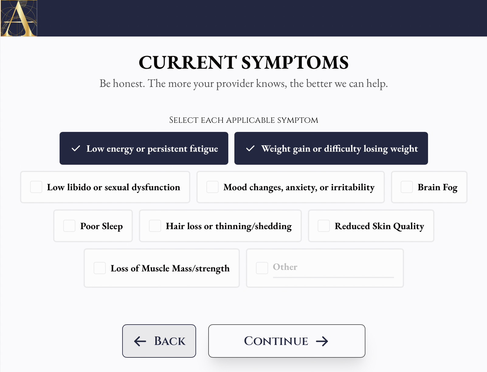
Online Assessment
Complete a brief intake covering your health history, current symptoms, and what you are looking to address.

Lab Testing
Select one of our blood work panels and complete your draw at a Quest location near you, or send us labs from the last 3 months. At-home draws are available for an added fee.

Provider Consultation
Meet with your licensed medical provider for a live video or phone consultation where they review your results, explain what they mean, and build your protocol with you.

Start Treatment
If treatment is clinically appropriate, your provider prescribes through one of our licensed partner pharmacies and your medications ship directly to your home.
Learn More
Expert insights and educational resources
No blog posts selected. Please select posts in the element settings.
Common questions
Can't find what you're looking for?
